This project began when a science center requested a 4ft tall interactive brain exhibit. Although it was ultimately cancelled, I decided to carry on by creating a physical, hands-on brain and combining it with a virtual component for a unique and educational experience.
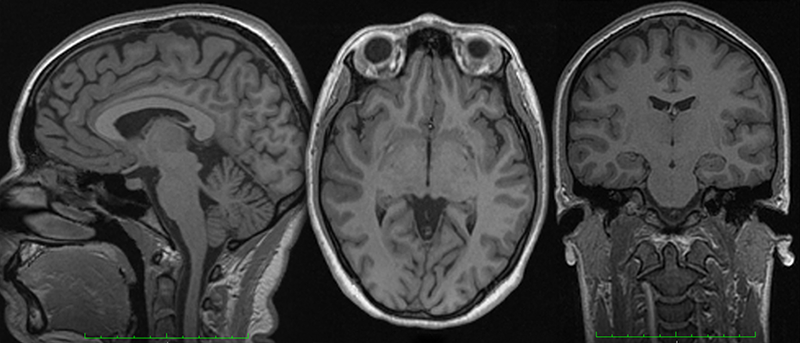
First, I participated as a control in a medical study in order to obtain MRI images of my brain:
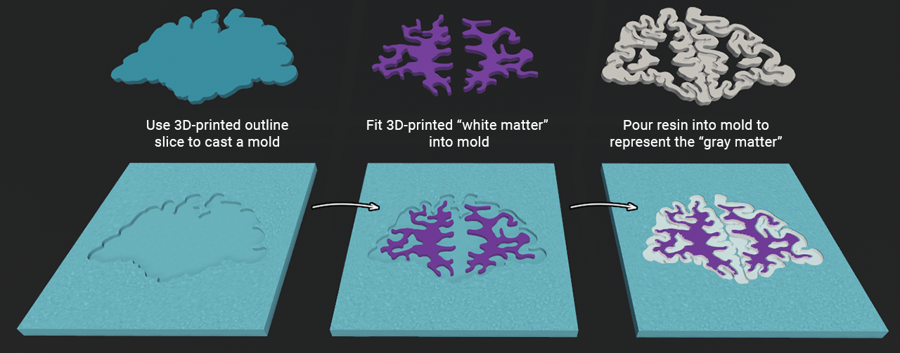
After selecting 26 equidistant slices along the coronal cross section, I created 3D models of the slice outline, (inner) white matter, and (outer) gray matter.
- Each slice outline was 3D printed and used to cast a rubber silicone mold.
- The white matter was 3D printed in a bright color (purple) for visibility, and fit into the mold.
- Lastly, resin was poured into the mold. It settled and solidified around the white matter to represent gray matter, but still ensure some visibility behind it.
I came up with a few ideas of how the brain slices should be arranged and held, keeping in mind that multiple people might interact with it at the same time, and might not know how to put it back together. I decided on creating wooden platforms of varying sizes for each slice, held by a large block with slats that ensure each slice has a perfectly fitting counterpart.
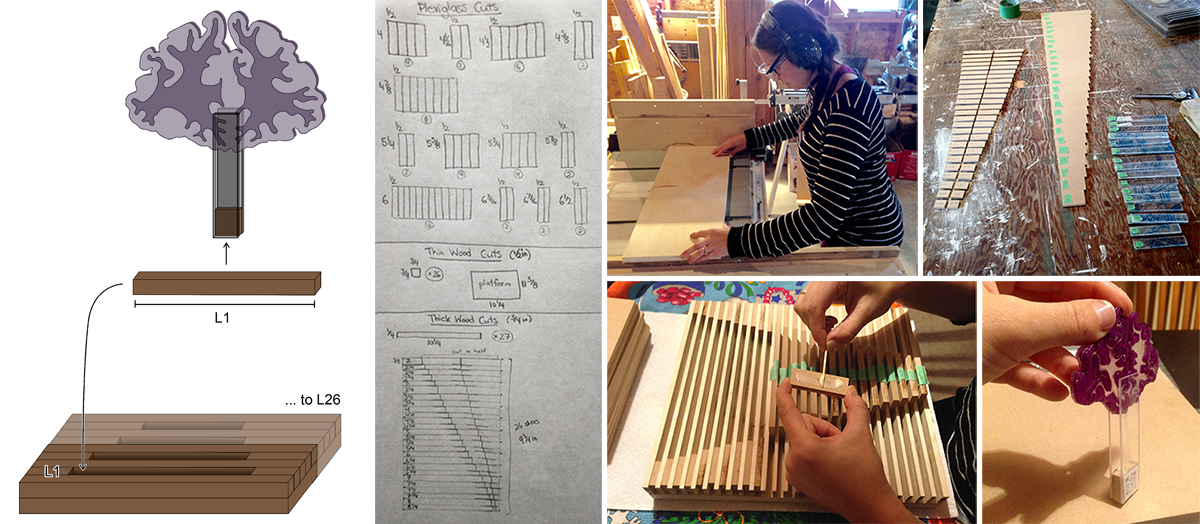
The final result was physically to scale with my own brain, but the brain was stretched along one axis due to the width of each slice. The experiments I conducted with laser cut acrylic slices might have mitigated that issue, but that is a project for the future - I still had a virtual component to add.

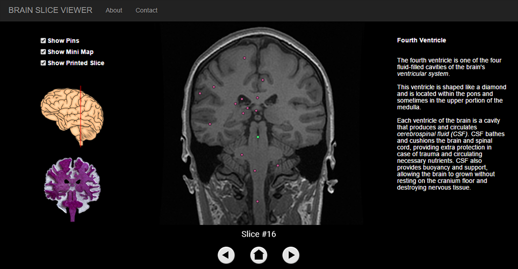
To finish off my tool, I wanted to build a web app with an informative page for every slice. The website is meant to be accessed through QR codes located at the base of each physical slice.

To understand the relative location of the slice in the brain, a saggital view of the brain was included. To connect the app to the physical hands-on exhibition, a matching icon of the 3D printed slice was added as well.
Finally, the original MRI scan would be front and center, with clickable pins for major components visible in the slice. Every slice has pins that were verified by a medical professional to ensure the information was accurate.
The intended goal was to eventually provide professors the ability to customize the pins and information presented on the web app for their class, or allow museums or other public spaces to include the information they wanted their visitors to focus on.